Bioimaging and Biocomputing Facility TLL
OVERVIEW
MICROSCOPY.TLL.ORG.SG RANKINGS

Date Range

Date Range

Date Range
LINKS TO BUSINESS
Click banner for more info and registration details. Temasek Life Sciences Laboratory, Singapore and Anhui Rice Research Institute, China Collaborate on Rice Research and Manpower Training, 6 March 2015. Temasek Life Sciences Laboratory Appoints Prof Yu Hao as Executive Director, 31 December 2014.
WHAT DOES MICROSCOPY.TLL.ORG.SG LOOK LIKE?
MICROSCOPY.TLL.ORG.SG HOST
BOOKMARK ICON

SERVER OPERATING SYSTEM
I discovered that this website is utilizing the Apache server.TITLE
Bioimaging and Biocomputing Facility TLLDESCRIPTION
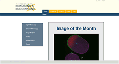
Objectives and Filter Sets. Request for HPC Account. TLLs Galaxy and UCSCGB. Image of the Month. Invasive hyphal growth of Magnaporthe oryzae in live rice cells. Conidia of the M. oryzae were incubated on the rice sheath. Two days after inoculation, the fungal hyphae invaded a neighboring rice cell through plasmodesmata arrows. The image was taken on the spinning disk confocal, acquired with 60X1.4 objective. Taken by Yang Fan.CONTENT
This site microscopy.tll.org.sg has the following on the web page, "Invasive hyphal growth of Magnaporthe oryzae in live rice cells." Our analyzers observed that the website also said " oryzae were incubated on the rice sheath." The Website also stated " Two days after inoculation, the fungal hyphae invaded a neighboring rice cell through plasmodesmata arrows. The image was taken on the spinning disk confocal, acquired with 60X1."MORE WEBSITES
Arkansas Nano and Bio Materials Characterization Facility. Nanoscale instruments and expertise for campus and the state. 2015, The University of Arkansas.
Please note that links from this site point to an updated Microscopy Core Website. The UCSD Health Sciences Microscopy Core is a state-of-the-art imaging core facility that serves the needs of laboratories in and outside of the UCSD School of Medicine. The Core strives to promote interdisciplinary, collaborative research among the local research community.
Darr; Skip to Main Content. CoolLED pE - 300 lite. CoolLED pE - 300 ultra. Two Chambered McMaster slide with imprinted grid. Three Chambered McMaster slide with imprinted grid.
Hans Ris seminar series with Erik M. In vivo multimodal bioimaging with Xuemei Wang. Quantitative 3D imaging using light sheet with Reto Fiolka. 9th Annual MERI vision science symposium with Steven Seitz. Frontiers in vision research with James Tahara Handa. Hitachi 3400 Variable Pressure SEM. CAMECA SX Five FE Electron Probe Microanalyzer. Nikon Andor Spinning Disk Confocal. Zeiss Auriga Focused Ion Beam FE SEM. Amnis Image Stream Mark II.